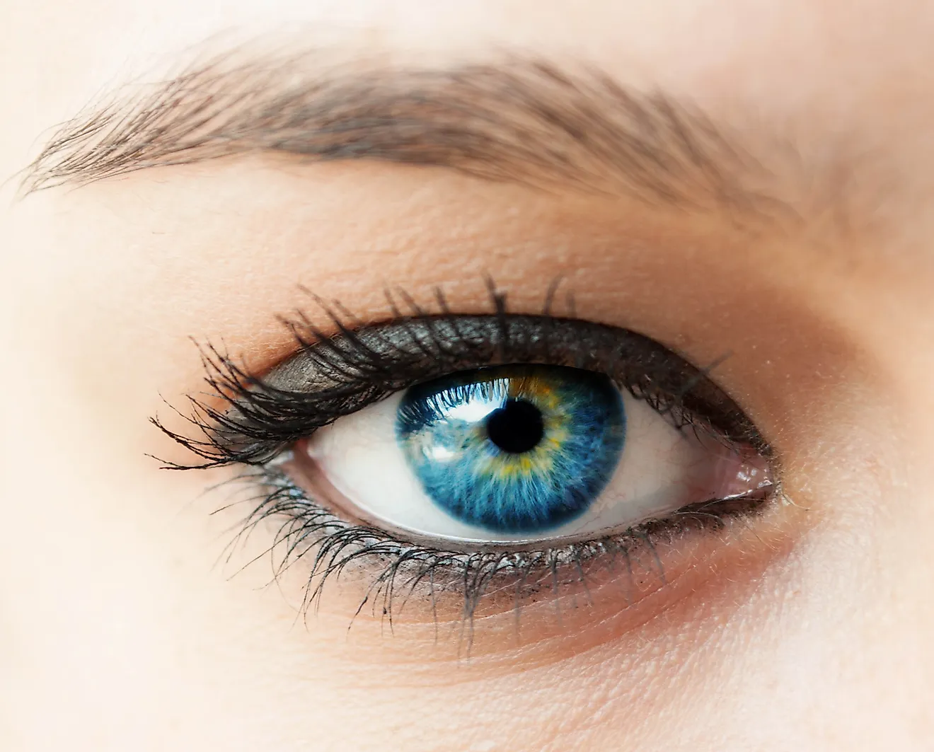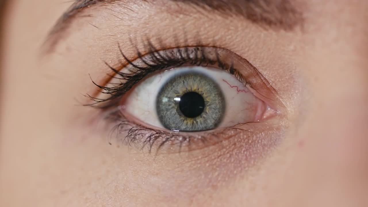
This is the ring that later forms the cornea and the eye's lens.
Human eyeballs skin#
Think of them as one of the earliest and simplest phase in biological eye formation.įor this study, Nishida's colleagues induced the formation of these proto-eyes from rabbit skin cells, and then pulled out a chunk of forming cells from the third of the proto-eye's four rings. Essentially, these proto-eyes amount to four simple rings of different cell types that later transform into different parts of the eye, like the retina or lens. The researchers kick-start the formation of this proto-eye by growing iPS cells in a petri dish lined with the right combination of proteins and other molecules. This new team discovered they could coerce these iPS cells into forming a simple proto-eye, from which they could harvest a bounty of different eye tissues. This discovery won two researchers, Shinya Yamanaka and Sir John Gurdon, the 2012 Nobel Prize in Physiology or Medicine the engineered cells are called induced pluripotent stem cells, or iPS.

In 2006, scientists found they could create fully fledged stem cells from ordinary child or adults cells like blood or skin with just a few simple DNA tweaks.

Nishida's new research builds upon a recent breakthrough in stem cell technology. He believes that within the next three years, we will be able to run trials to repair disease- or injury-damaged human corneas. "We are now in the position to initiate first in-human clinical trials of anterior eye transplantation to restore visual function," Nishida writes in today's Nature paper. Don't throw away your glasses (or eye patch) just yet, but human trials are up next. The research is published today in the journal Nature.

In a preliminary trial, the Japanese researchers cultured and grew sheathes of rabbit cornea-the transparent cover of the eye- that restored sight in blind rabbits born without fully-grown corneas. Using their new method, Nishida's team can grow retinas, corneas, the eye's lens, and more. No 11-cis isomer was formed under these conditions.A breakthrough in stem cells just brought us much closer to lab-grown human eyeballs.īiologists led by Kohji Nishida at Osaka University in Japan have discovered a new way to nurture and grow the many separate tissues that make up the human eyeball, and the scientists need only a small sample adult skin to build them all. The all-trans configuration was mainly retained during uptake and esterification, although some isomerization to 13-cis also occurred. Intact RPE-Ch or isolated RPE cells esterified exogenous all-trans-3H2-retinol to the same fatty acids in roughly the same proportions as in the endogenous stores. Variable, usually small proportions of 13-cis retinyl esters were also present.

The ratio of 11-cis isomer over the all-trans averaged 1.52 +/- 0.48 (n - 11). A small amount of oleate was also detected. A preponderance of the vitamin A in the eye was esterified (98.3% in the RPE-Ch, 79.3% in the retina) and consisted principally of stearate and palmitate in the ratio of 1:4.8. By measuring rhodopsin regeneration in retinal homogenates incubated with 11-cis retinal, we estimated that the amount of vitamin A in the RPE-Ch of fully dark-adapted eyes would represent 2.5 mole equivalents of the retinal rhodopsin, a value similar to that found in the frog. The vitamin A extracted from the retina was 15.3% of that in the corresponding RPE-Ch. There was no evidence for significant losses during the interval between death and enucleation or during subsequent storage at 4 degrees C. The vitamin A concentration in the pigment epithelium-choroid (RPE-Ch) was the highest observed for human non-liver tissue and amounted to 7.9 +/- 4.3 nmol/eye (n = 28), or 10.4 +/- 7.1 microgram/gm (n = 27). The amount, distribution, and composition of vitamin A stored in the eyes of 29 postmortem donors was determined by a combination of techniques, including high-pressure liquid chromatography.


 0 kommentar(er)
